X-ray imaging is a widely utilized medical diagnostic tool that helps healthcare professionals visualize the internal structures of the body. By using X-rays, which are a form of electromagnetic radiation, images are produced showing various tissues, bones, and organs. This method is quick, non-invasive, and offers valuable insights into a patient's health. Whether it’s for diagnosing fractures, identifying infections, or screening for diseases, understanding how X-ray imaging works is foundational for effective analysis. In this section, let’s dive deeper into what X-ray imaging really entails.
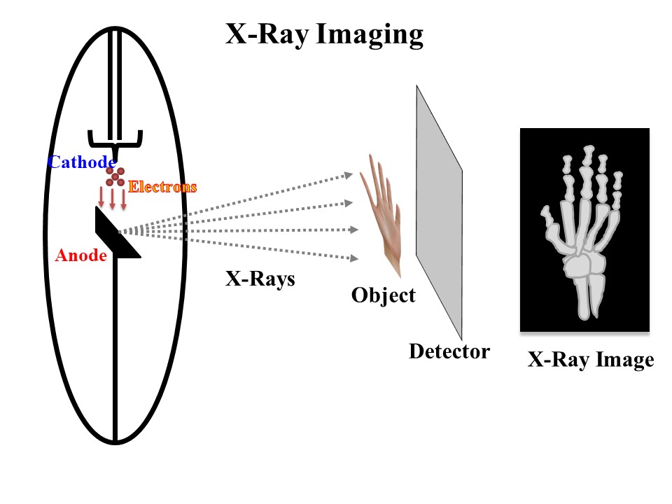
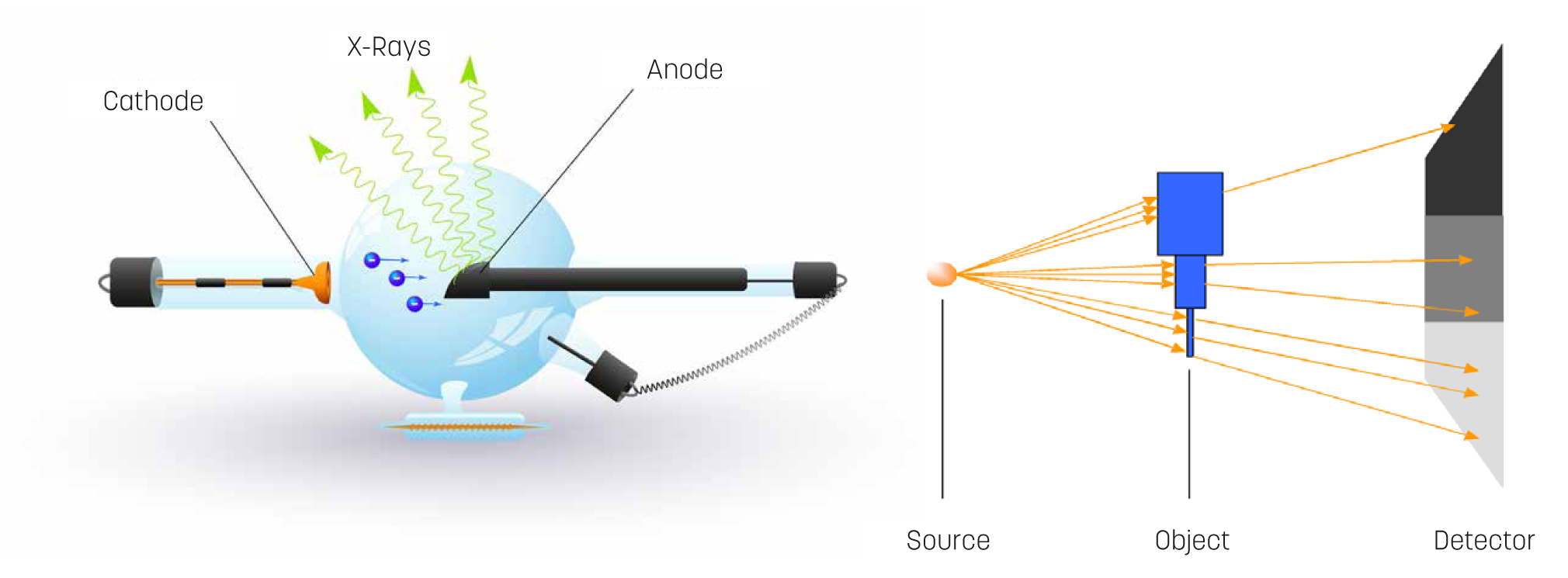
At its core, X-ray imaging involves directing X-ray beams at a patient, which then pass through the body and are captured on a detector or film. The resulting images appear in shades of black, white, and gray—indicating different levels of tissue density.
To break it down further:
- X-Ray Production: X-rays are generated when high-energy electrons from an X-ray tube collide with a metal target, usually tungsten. This interaction produces X-rays that exit the tube.
- Radiolucent vs. Radiopaque: Different tissues absorb X-rays to varying degrees. For example, bones, being denser, appear white on the X-ray, while softer tissues like muscles and fat show up in shades of gray.
- Types of X-Ray Images: There are various forms, including conventional X-rays, computed tomography (CT) scans, and fluoroscopy. Each serves a unique purpose based on the detail required.
With a solid understanding of how X-rays work, healthcare professionals can make informed decisions during analysis, leading to better patient outcomes.
The Importance of X-Ray Analysis
The ability to analyze X-ray images effectively is crucial in medical diagnostics. X-ray analysis not only assists in identifying health issues but also plays a role in treatment planning and monitoring progress. It's the bridge between a physical examination and a diagnosis, providing visual evidence that informs clinical decisions. Let’s explore why X-ray analysis is so significant.
Here are some key points illustrating the importance of X-ray analysis:
- Early Detection: X-rays can reveal conditions like fractures, tumors, or infections before they become critical. Early detection often results in better treatment outcomes.
- Guiding Treatment: By accurately diagnosing a condition through X-ray analysis, healthcare providers can recommend the most effective treatment options tailored to the patient's specific needs.
- Monitoring Progress: X-ray images taken over time can help track the effectiveness of a treatment plan, allowing for adjustments if necessary.
| Key Benefits of X-Ray Analysis | Examples |
|---|---|
| Accurate Diagnosis | Detecting lung infections or tumors |
| Non-Invasive | Assessing joint health without surgery |
| Cost-Effective | Reducing the need for more expensive diagnostic procedures |
In summary, effective X-ray analysis is not just a skill; it’s a critical component of successful medical care. Knowing how to interpret these images properly is essential for accurate diagnoses and effective treatment strategies, ultimately leading to improved patient outcomes.
Basic Principles of X-Ray Technology
X-rays have revolutionized the field of medical imaging, allowing healthcare professionals to peep inside the human body without any invasive procedures. At the core of X-ray technology lies the simple principle of exposing a patient's body to a controlled amount of radiation, which helps in creating images of internal structures.
To understand how this works, it’s essential to grasp a few key concepts:
- Radiation: X-rays are a type of electromagnetic radiation, similar to light but with much higher energy. This allows them to penetrate through soft tissues, which is why bones, which are denser, show up clearly.
- Film/Detector: When X-rays pass through the body, they are either absorbed or transmitted, depending on the density of the materials they encounter. This difference is captured on a film or digital detector, resulting in an image that highlights various structures.
- Contrast Agents: Sometimes, to enhance the visibility of certain areas, contrast agents may be introduced. These substances absorb X-rays differently than surrounding tissues, making specific organs more visible.
It’s important to remember that, while X-rays are invaluable, they do involve exposure to radiation. While the doses are typically low and considered safe, understanding how this technology operates offers valuable context for its usage in medical assessments.
Steps to Analyze X-Ray Images
Analyzing X-ray images requires a systematic approach to ensure accurate interpretation. Here’s a simple step-by-step guide for you:
- Step 1: Obtain a Quality Image
Before diving into analysis, ensure that the X-ray is of high quality. Look for clarity, proper exposure, and correct positioning of the patient during the shot.
- Step 2: Review Patient History
Understanding the patient’s medical history and symptoms is crucial. This context helps in narrowing down potential issues that may be visible on the X-ray.
- Step 3: Systematic Examination
Adopt a systematic approach while analyzing the image. A common practice is to examine from one anatomical area to another, ensuring nothing is overlooked. Make observations about:
- Bone structure and integrity
- Presence of fractures or abnormalities
- Soft tissue characteristics
- Step 4: Look for Asymmetry
Compare both sides of the body. Asymmetric findings can often indicate underlying issues that need further investigation.
- Step 5: Take Notes
Document your findings in a structured manner. This allows you to discuss specifics with colleagues and helps in future reference.
- Step 6: Seek a Second Opinion
If you have doubts or the finding is complex, don’t hesitate to consult with another professional. Collaboration can lead to better patient outcomes.
By following these steps, you'll be able to analyze X-ray images more effectively, leading to accurate diagnoses and better patient care.
5. Common Conditions Identified in X-Ray Images
When you think about X-ray imaging, you might picture a clear look at broken bones or dental issues. However, X-rays can reveal much more than that! Here’s a rundown of some common conditions that healthcare professionals often identify through X-ray images:
- Fractures: One of the most common reasons for an X-ray is to diagnose fractures. Whether it's a simple wrist fracture or a complex broken leg, X-rays provide a clear image of bone structure.
- Arthritis: X-rays can show changes in the joints, such as swelling or bone spurs, helping to diagnose various types of arthritis.
- Pneumonia: Chest X-rays can reveal fluid in the lungs, helping doctors diagnose and treat pneumonia effectively.
- tumors: Certain X-ray techniques can help spot abnormal growths in various organs, making them important tools for cancer screening.
- Bone Loss: Conditions like osteoporosis can cause reduced bone density, which X-rays can detect over time.
By recognizing these common conditions, both healthcare providers and patients can have a clearer understanding of what an X-ray can reveal. Remember, diagnosing a condition through an X-ray is just one part of the overall assessment; often, additional testing is required for a full diagnosis!
6. Tools and Software for X-Ray Image Analysis
Analyzing X-ray images effectively goes beyond just looking at the pictures; it often requires sophisticated tools and software that enhance the diagnostic process. Here’s a look at some of the standout options:
| Tool/Software | Description | Key Features |
|---|---|---|
| PACS (Picture Archiving and Communication System) | A medical imaging technology used for storing, retrieving, presenting, and sharing images. | Remote access, integration with other medical systems, and security features. |
| CAD (Computer-Aided Detection) | Software that identifies potential abnormalities in X-ray images to aid radiologists. | Automated detection, real-time analysis, and support for various imaging modalities. |
| Image Enhancement Tools | Software that improves the quality and clarity of X-ray images for better diagnostic accuracy. | Contrast adjustment, sharpening, and noise reduction capabilities. |
| AI-Powered Analysis Tools | Advanced software utilizing artificial intelligence to assist in diagnosing conditions. | Machine learning algorithms, predictive analytics, and large data training sets. |
Using these tools streamlines the diagnostic process, increases accuracy, and ultimately leads to better patient care. The right software not only supports medical professionals in their analysis but also promotes collaboration and data sharing for improved outcomes.
7. Best Practices for Accurate X-Ray Interpretation
When it comes to interpreting X-Ray images, accuracy is vital for effective diagnosis and treatment planning. Here are some best practices that radiologists and healthcare professionals should keep in mind:
- Know the Radiographic Principles: Understand the basic principles of radiography, including contrast, density, and exposure settings. This knowledge will help you make more informed observations.
- Systematic Approach: Adopt a structured method for analysis. Consider using the ABCDEs of X-Ray interpretation (Alignment, Bones, Cartilage, Detection of soft tissues, and Everything else), ensuring you don’t overlook any crucial details.
- Compare with Previous Images: If available, always compare with earlier X-Rays of the same patient. This can be invaluable for detecting changes over time.
- Focus on the Clinical Context: Always consider the patient’s symptoms, history, and other clinical findings. This context helps in narrowing down potential issues and aids in prioritizing findings.
- Consult with Colleagues: Don’t hesitate to collaborate with others. Discussing findings with colleagues can bring fresh perspectives or highlight aspects you may have missed.
- Continual Education: Keep learning! Attend workshops, webinars, and courses about the latest in radiology and imaging techniques. Staying updated helps enhance your skills.
By adhering to these practices, healthcare professionals can significantly improve their accuracy in interpreting X-Ray images, leading to better patient outcomes.
8. Conclusion: Key Takeaways for X-Ray Image Analysis
In closing, analyzing X-Ray images is a skill that combines technical knowledge, clinical insight, and continual education. Here are some key takeaways to keep in mind:
- Importance of Training: Proper training and education are crucial for mastering X-Ray interpretation. This can’t be emphasized enough.
- Use of Technology: Embrace advancements in imaging technology and software that enhance diagnostic capabilities, but don’t rely solely on them.
- Patient-Centered Approach: Always keep the patient’s broader clinical scenario in mind; it provides context that can shape your interpretation of the image.
- Team Collaboration: Engaging with a multidisciplinary team can broaden the diagnostic reach and help in confirming findings.
- Practice Makes Perfect: Regularly reviewing images and participating in case study discussions can refine your skills over time.
By following these guidelines, radiologists and healthcare professionals can enhance their capacities for accurate X-Ray image analysis, ultimately leading to improved healthcare delivery and patient satisfaction.