Ultrasound imaging is a powerful tool used in medical settings to visualize internal organs and tissues. It uses high-frequency sound waves to create images of the body. This non-invasive technique is commonly used during pregnancy but also helps diagnose various health conditions. One of the best things about ultrasound is that it doesn't involve radiation, making it a safe option for patients.
Understanding Ultrasound Images

Reading ultrasound images can seem daunting at first, but with some basic knowledge, it becomes easier. Ultrasound images are essentially pictures created by sound waves bouncing off body structures. These
- Echo Patterns: Dark areas indicate fluid, while light areas suggest solid tissues.
- Transducer Position: The position of the ultrasound probe affects the image, so it's essential to know where the probe is placed.
- Fetal Imaging: In pregnancy, the images are used to assess the baby's growth and well-being.
Getting familiar with these basics helps in interpreting ultrasound images with more confidence.
Common Types of Ultrasound Images

There are several types of ultrasound images used for different medical purposes. Understanding these types can help you know what to expect during an ultrasound. Here are some of the most common ones:
| Type of Ultrasound | Description |
|---|---|
| 2D Ultrasound | This is the standard ultrasound, providing flat, two-dimensional images of organs. |
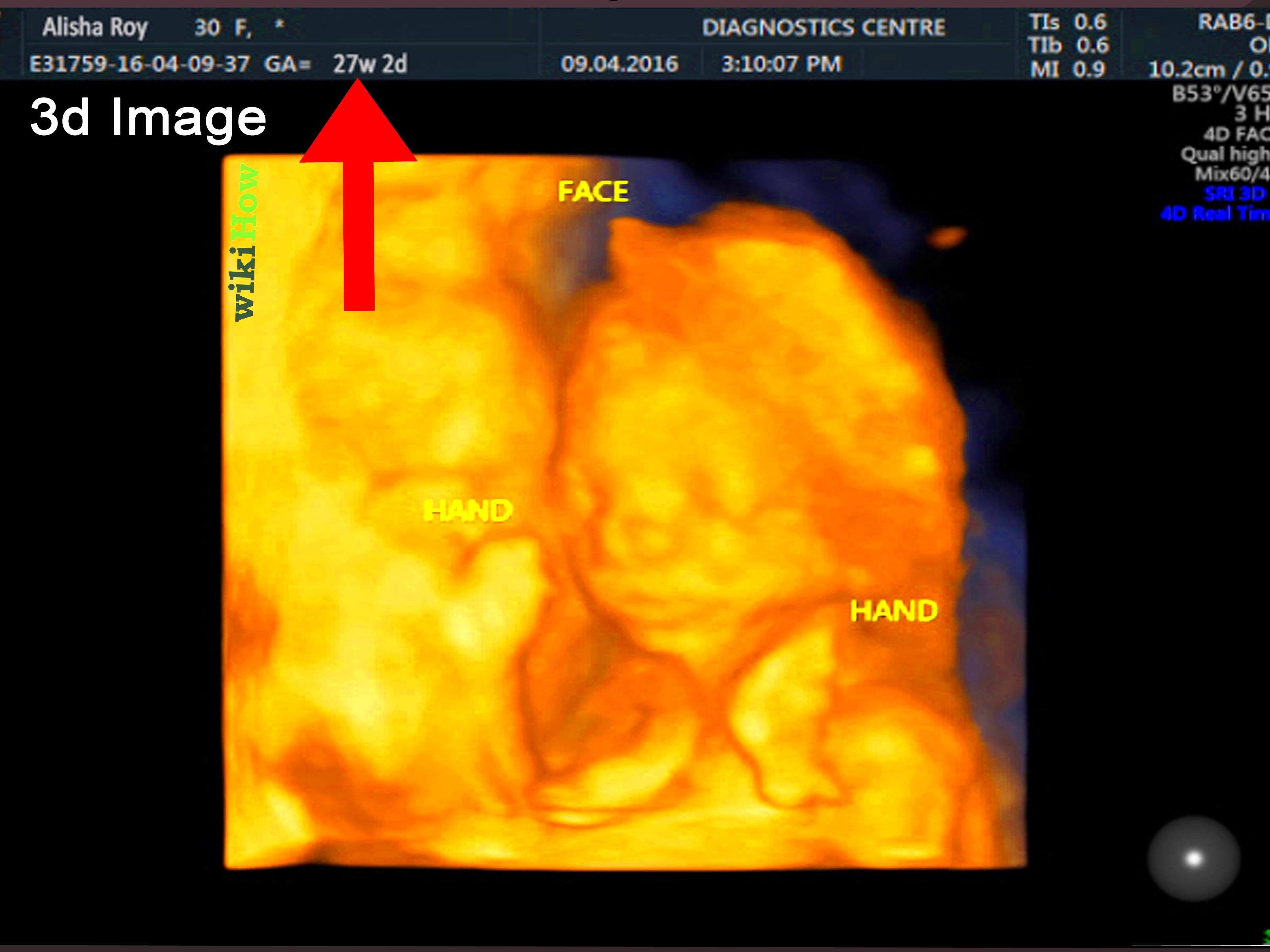
| 3D Ultrasound | This type gives a three-dimensional view, often used in prenatal imaging to provide a more detailed look at the baby. |
| 4D Ultrasound | Similar to 3D but adds motion, allowing for real-time imaging of the fetus. |
| Doppler Ultrasound | This type measures the flow of blood in the body and can detect abnormalities in blood circulation. |
Each type has its specific uses, making ultrasound a versatile imaging technique in healthcare.
Steps to Read Ultrasound Images

Reading ultrasound images may seem complex, but breaking it down into steps can simplify the process. Here are the essential steps to help you interpret ultrasound images effectively:
- Preparation: Before reading the images, ensure you have background information on the patient and the reason for the ultrasound.
- Initial Review: Start by looking at the overall image quality. Check for clarity, orientation, and any artifacts that might affect interpretation.
- Identify Planes: Recognize the imaging planes, such as transverse, longitudinal, and oblique. Each plane offers a different perspective on the structures being examined.
- Focus on Anatomy: Familiarize yourself with normal anatomy relevant to the exam. Knowing what to look for helps in spotting abnormalities.
- Look for Pathologies: Observe any unusual patterns or structures. Common signs include cysts, masses, or changes in organ shape or size.
- Compare with Previous Studies: If available, compare the current images with previous ultrasounds for changes or developments.
- Consult Resources: Use textbooks or online resources to clarify any doubts about the structures or findings.
Following these steps can enhance your confidence and accuracy when interpreting ultrasound images.
Identifying Key Structures in Ultrasound Images
Being able to identify key structures in ultrasound images is crucial for accurate interpretation. Each organ and tissue type has unique characteristics that help differentiate them. Here are some common structures and tips to identify them:
- Kidneys: Look for a bean-shaped structure, usually located in the upper abdominal area. They appear hypoechoic (dark) against the surrounding tissue.
- Liver: The liver is typically larger and appears more echogenic (lighter) than the kidneys. It is located in the right upper quadrant.
- Gallbladder: This is a pear-shaped structure near the liver, often seen as an anechoic (dark) area when filled with bile.
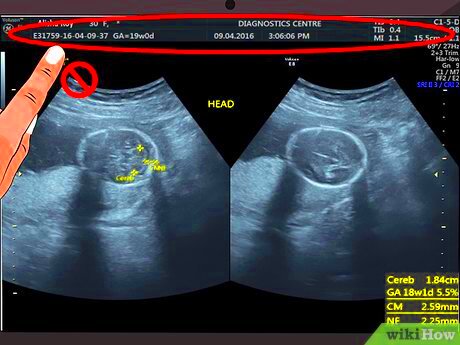
- Fetus: In pregnancy scans, a fetus can be recognized by its outline and distinct features like limbs and the head. The position of the fetus also offers clues.
Having a mental map of these structures and their typical appearances makes it easier to identify them during an ultrasound exam.
Common Challenges in Reading Ultrasound Images
Even experienced professionals can face challenges when reading ultrasound images. Understanding these common issues can help you navigate them more effectively:
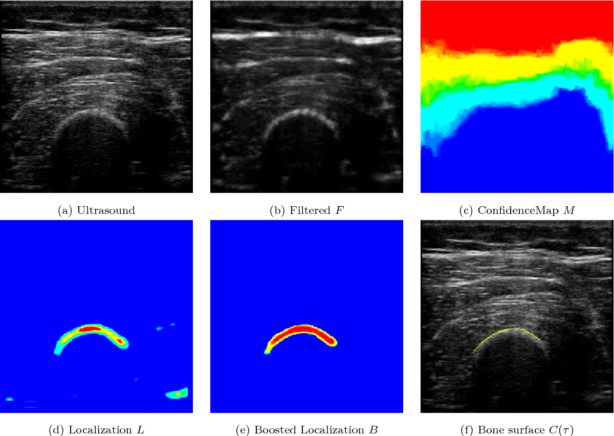
- Image Quality: Poor quality images can obscure important details. Factors like patient movement, inadequate transducer contact, or equipment settings can affect clarity.
- Variability in Anatomy: Individual differences in anatomy can lead to confusion. What is normal for one person may not be for another.
- Artifacts: Artifacts are false images that can appear on ultrasound due to various factors, such as shadowing or enhancement. Recognizing these helps avoid misinterpretations.
- Limited Experience: Lack of experience can make it difficult to differentiate between normal variations and pathological findings. Continuous learning is essential.
- Emotional Factors: If the ultrasound is for a sensitive situation, like pregnancy complications, emotions can cloud judgment and lead to misinterpretation.
Awareness of these challenges can prepare you for a more effective and confident approach to reading ultrasound images.
Tips for Improving Image Interpretation Skills
Improving your ultrasound image interpretation skills takes practice and dedication, but with the right tips, you can enhance your confidence and accuracy. Here are some strategies to help you on this journey:
- Practice Regularly: The more images you review, the better you will get. Make it a habit to study different ultrasound images regularly.
- Join Workshops: Participating in workshops or training sessions can provide hands-on experience and insights from experts in the field.
- Study Anatomy: A strong foundation in human anatomy is essential. Familiarize yourself with normal anatomical variations to recognize abnormalities more easily.
- Use Reference Materials: Keep textbooks or online resources handy. They can be invaluable for double-checking your findings and learning more about specific conditions.
- Work with Mentors: If possible, seek feedback from more experienced colleagues. They can offer tips and help you identify areas for improvement.
- Simulate Real Cases: Engage in case studies or simulations that mimic real-life scenarios. This practice can sharpen your analytical skills.
By implementing these tips, you can steadily improve your ultrasound image interpretation skills, making the process more intuitive and efficient.
FAQ about Reading Ultrasound Images
Here are some frequently asked questions about reading ultrasound images. These can help clarify common concerns and provide more insights:
| Question | Answer |
|---|---|
| What is the main purpose of ultrasound imaging? | Ultrasound imaging is primarily used to visualize internal organs, monitor fetal development, and diagnose various medical conditions. |
| Is ultrasound safe? | Yes, ultrasound is a safe, non-invasive procedure that does not use ionizing radiation, making it suitable for various patients, including pregnant women. |
| How long does an ultrasound exam take? | The duration of an ultrasound exam can vary, but it typically lasts between 20 to 60 minutes, depending on the complexity of the study. |
| What if I see something unusual on the ultrasound? | If you notice something abnormal, it's important to consult a medical professional for further evaluation and possible additional testing. |
These FAQs can help ease concerns and provide a better understanding of ultrasound imaging.
Conclusion on Ultrasound Image Reading
Reading ultrasound images is a valuable skill that combines knowledge, practice, and intuition. By following the steps outlined and employing tips to improve your interpretation skills, you can enhance your confidence in this area. Understanding the common types of ultrasound images, identifying key structures, and being aware of challenges are essential parts of this process. As you gain experience, you will become more adept at recognizing both normal and abnormal findings, ultimately contributing to better patient care. Remember, continuous learning and practice are the keys to mastering ultrasound image reading.

 admin
admin


