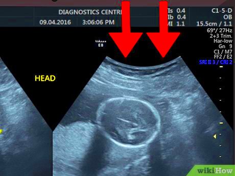
Ultrasound imaging, also known as sonography, is a medical procedure that uses high-frequency sound waves to create images of the inside of the body. Unlike other imaging techniques such as X-rays or MRIs, ultrasound doesn’t use radiation. This makes it a safer and non-invasive option for patients, especially in obstetrics and gynecology. It is commonly used to monitor pregnancy, examine internal organs, and guide certain medical procedures. Understanding the basics of ultrasound imaging can help you make sense of the images and interpret the results accurately.
How Ultrasound Images Are Created

Ultrasound images are generated using a device called a transducer, which sends out high-frequency sound waves. When these waves hit an object or tissue inside the body, they bounce back, or echo. The transducer then detects these echoes and sends them to a computer, which processes the signals and converts them into an image. The computer interprets the time it takes for the sound waves to return, the intensity of the echoes, and their direction to create a visual representation of the tissues or organs being examined.
Key steps in creating an ultrasound image include:
- Sound Wave Emission: The transducer emits sound waves into the body.
- Echo Reception: The sound waves bounce off tissues and return to the transducer.
- Image Creation: A computer processes the echoes and forms an image.
These images can appear in various formats, such as black-and-white or color Doppler images, depending on the type of ultrasound and the area being examined.
Key Components of an Ultrasound Image

Understanding the key components of an ultrasound image can make it easier to interpret what you’re looking at. There are several important aspects to consider when examining an ultrasound image:
- Brightness and Contrast: The brightness of the image indicates how dense or fluid-filled the tissue is. Darker areas often represent fluid-filled spaces (like cysts), while lighter areas are denser structures, such as bones or tissues.
- Resolution: The resolution of an ultrasound image refers to the level of detail that can be seen. High-resolution images provide clearer details, which are crucial for accurate diagnosis.
- Measurement: Ultrasound images often include measurements, particularly when tracking the growth of a fetus or the size of an organ. These measurements help doctors make assessments about the health of the patient.
Here's a quick breakdown of common terms you’ll encounter in an ultrasound image:
| Term | Description |
|---|---|
| Hypoechoic | Areas that appear darker on the image, often indicating less dense tissue. |
| Hyperechoic | Brighter areas on the image, typically indicating denser tissues like bone. |
| Anechoic | Completely black areas, often indicating fluid-filled structures like cysts or blood vessels. |
By understanding these components, you can better interpret the images and the health information they provide.
What to Look for in an Ultrasound Image

When looking at an ultrasound image, there are several things to focus on to better understand the condition or state of the area being examined. Ultrasound images can be a bit overwhelming at first, but with the right knowledge, you can start to pick up on key features that provide valuable insights. The main things to look for include the structure, size, shape, and texture of the tissues or organs being visualized. It’s also important to note any unusual masses, fluid buildup, or areas that may seem abnormal. Here are the primary things to focus on:
- Size and Shape: Ensure the size and shape of organs or structures are normal. For example, an enlarged organ might suggest an underlying issue.
- Texture: Pay attention to the texture of the tissues. Uniform textures are usually normal, while irregular textures could signal a problem.
- Presence of Masses: Look for any masses, lumps, or abnormal growths, which can indicate tumors or cysts.
- Fluid Buildup: Check for areas of fluid accumulation, which might point to conditions like edema or abscesses.
Remember that a clear understanding of what the image should look like in normal conditions will help you better identify anything out of the ordinary. Sometimes the images need to be examined alongside other diagnostic tests to get a complete picture.
Common Types of Ultrasound Images and Their Purposes
There are various types of ultrasound exams, each designed to visualize different aspects of the body. The purpose of the ultrasound and the area being examined determines the type of image produced. Below are the most common ultrasound types and what they’re used for:
- 2D Ultrasound: The most common type, creating flat, two-dimensional images of internal structures. This is typically used during pregnancy to monitor the baby’s development.
- 3D Ultrasound: A more advanced technique that creates a three-dimensional image. It’s used in obstetrics to provide more detailed images of the baby.
- 4D Ultrasound: Similar to 3D but with the added ability to show movement. It’s often used to watch the baby’s movements in the womb.
- Doppler Ultrasound: This type assesses blood flow by measuring the frequency of the sound waves bouncing off moving blood cells. It's commonly used to check heart conditions or blood clots.
- Elastography Ultrasound: A special type of ultrasound that measures tissue stiffness, often used to evaluate liver diseases or cancers.
Each type of ultrasound serves a unique purpose and can offer crucial information depending on the area of concern. For example, 2D ultrasound is most commonly used in early pregnancy to confirm fetal heartbeat, while Doppler ultrasound is essential in assessing vascular conditions like blood clots or blocked arteries.
Challenges in Interpreting Ultrasound Images
Interpreting ultrasound images can be tricky, especially for beginners. While ultrasound is a valuable diagnostic tool, there are some challenges that may make the images harder to understand. One of the main difficulties is that the quality of ultrasound images can vary based on factors such as the skill of the technician, the type of equipment used, and the condition of the patient. Here are some challenges to keep in mind:
- Image Quality: Poor image resolution can make it difficult to differentiate between structures, leading to possible misinterpretation. In some cases, the image may not be clear enough to detect small abnormalities.
- Patient Factors: Obesity, the position of the organs, or air/gas in the intestines can interfere with the clarity of the ultrasound image. These factors can make it harder for the sound waves to pass through the body effectively.
- Overlapping Structures: Ultrasound images often display overlapping or similar-looking structures, which can confuse the interpretation. Distinguishing between normal and abnormal tissues can be challenging without the right experience.
- Operator Skill: The skill of the person operating the ultrasound machine plays a big role in image clarity. An experienced technician will know how to adjust settings and angle the transducer for the best image quality.
Despite these challenges, ultrasound remains a highly effective imaging tool. However, for an accurate diagnosis, it’s crucial to combine ultrasound findings with other tests, a detailed medical history, and physical exams.
Tips for Beginners in Reading Ultrasound Images
Reading ultrasound images can seem challenging at first, but with a little practice and knowledge, you can start to feel more confident in interpreting them. Whether you're a student, a healthcare professional, or just someone interested in the process, it's important to understand the basics and focus on key areas in the image. Here are some helpful tips to get you started:
- Familiarize Yourself with Normal Anatomy: The more you know about the anatomy of the body and how it should appear on an ultrasound, the easier it will be to spot abnormalities. Start with common organs and structures like the liver, kidneys, and heart.
- Learn the Terminology: Ultrasound images come with their own set of terms, such as "hypoechoic," "hyperechoic," and "anechoic." Understanding these terms will help you interpret the images more accurately.
- Practice with Examples: The best way to improve your skills is by practicing. Look at as many ultrasound images as possible, even if you're not sure what you're seeing. Over time, patterns and key features will become clearer.
- Focus on Key Features: Pay close attention to the size, shape, and texture of organs. Identify fluid-filled areas, masses, or irregularities, as these can indicate underlying conditions.
- Ask for Guidance: If you're unsure about a particular image, don't hesitate to ask an experienced professional for guidance. Their input can be invaluable in helping you interpret complex images.
With these tips in mind, you can begin to read ultrasound images with more confidence. Remember, practice and patience are key to mastering this skill.
Frequently Asked Questions
If you're new to ultrasound imaging, you may have several questions about how the process works and what the images represent. Here are some of the most frequently asked questions to help you better understand ultrasound imaging:
- What does it mean if an area is "hypoechoic"? A "hypoechoic" area appears darker on the ultrasound image and typically indicates that the tissue is less dense, like fluid or certain types of soft tissue.
- Why do some ultrasound images look blurry or unclear? Several factors can affect the quality of an ultrasound image, including the skill of the technician, the patient's body type, and the presence of air or gas in the body.
- Can ultrasound images detect cancer? Ultrasound can help identify masses or tumors, but it can’t always determine whether they are cancerous. Further tests, like a biopsy or MRI, are needed for a more accurate diagnosis.
- Is ultrasound safe? Yes, ultrasound is generally considered very safe because it doesn’t use radiation, unlike X-rays or CT scans. It is non-invasive and can be used on pregnant women and infants without harm.
- Can ultrasound be used for all areas of the body? While ultrasound is effective for examining soft tissues like muscles, organs, and blood vessels, it is less effective for viewing areas like bones or the lungs.
If you have more specific questions or concerns, it’s always a good idea to consult with a healthcare professional who can guide you through the ultrasound process.
Conclusion
Reading ultrasound images may seem like a daunting task at first, but with a little practice and knowledge, it becomes much easier. Ultrasound is a powerful tool in modern medicine, offering a safe, non-invasive way to view internal structures and monitor health. By understanding the basics of how ultrasound works, the different types of images, and key components to look for, you’ll be better equipped to interpret these images effectively. Remember, practice is essential, so don’t be discouraged if you don’t get everything right at first. Over time, you’ll develop a sharper eye for detail, making ultrasound reading a valuable skill. If in doubt, always consult a professional for an accurate diagnosis.

 admin
admin


