Intraoral images are an essential part of modern dental care. These images are taken using specialized imaging equipment that captures detailed pictures of the inside of the mouth. Dentists use intraoral images to examine teeth, gums, and other oral structures, helping them identify issues that might not be visible during a regular exam. Intraoral images provide clarity and precision, making it easier to spot potential problems early and ensure that patients receive the right treatment. This technology is particularly useful for diagnosing conditions like cavities, gum disease, and even oral cancers.
For patients, understanding the purpose and benefits of intraoral images can help reduce any concerns about the process. These images play a key role in proactive dental care, ensuring healthier teeth and gums over the long term.
What is a Complete Series of Intraoral Images?

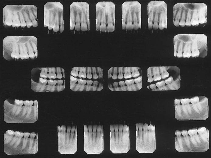
A complete series of intraoral images refers to a set of X-ray pictures that provide a full view of a patient’s mouth. This series usually includes multiple types of images, each focusing on a different part of the mouth. The goal is to capture a detailed, comprehensive view of both the teeth and surrounding tissues.
Typically, a complete series includes:
- Bitewing X-rays: These images show the upper and lower teeth in a bite position, helping dentists spot cavities between teeth.
- Periapical X-rays: These focus on individual teeth, showing the tooth and the bone structure surrounding it.
- Occlusal X-rays: These provide a broader view of the teeth and jaws, helping to identify issues like jaw fractures or developmental problems.
- Panoramic X-ray: A full view of the mouth, showing all the teeth, jaws, and sinuses in a single image.
When taken together, these images give a clear, complete picture of a patient’s oral health, allowing the dentist to diagnose any issues and create a treatment plan if necessary.
Also Read This: Converting Images to SSTV Scans
Why It is Important to Take a Complete Series
Taking a complete series of intraoral images is important for several reasons, primarily because it helps the dentist detect problems that might not be visible during a routine exam. Even with a thorough visual inspection, some issues may be hidden beneath the surface of the teeth or gums, and that’s where imaging technology comes in.
Here are a few key reasons why a complete series is essential:
- Early detection of dental problems: A complete series allows dentists to catch issues like cavities, tooth decay, or bone loss early, preventing them from progressing into more serious conditions.
- Accurate diagnosis: The images provide a detailed, clear view of both the teeth and the surrounding tissues, ensuring a more accurate diagnosis and treatment plan.
- Monitor oral health over time: A complete series taken at regular intervals can help dentists track changes in a patient’s oral health, allowing them to make adjustments to the treatment plan if needed.
- Personalized treatment plans: With a full view of the patient’s mouth, the dentist can offer more personalized care, focusing on the areas that need attention the most.
In short, a complete series of intraoral images ensures that dentists can provide the best possible care, helping patients maintain long-term oral health with early intervention and tailored treatment plans.
Also Read This: Cropping Images in Blender: A Tutorial
Factors Affecting the Frequency of Taking Intraoral Images
The frequency of taking intraoral images can vary based on several factors, each contributing to the decision-making process for both the dentist and the patient. In general, intraoral images are taken based on the patient’s unique oral health situation, medical history, and age. It's essential to consider these factors to determine the right timing for the imaging process.
Here are some key factors that influence how often intraoral images should be taken:
- Age: Children, for example, may require more frequent imaging as their teeth develop, while adults may need images less often, depending on their oral health.
- Oral health status: Patients with a history of cavities, gum disease, or other dental issues may need more frequent imaging to monitor their condition.
- Risk of disease: Individuals at a higher risk for oral health problems, such as those with diabetes or a history of oral cancer, might require more regular imaging.
- Routine dental checkups: Regular visits to the dentist help track changes in oral health and determine when imaging should be done. Dentists may adjust the frequency of images based on what they observe during exams.
- Specific concerns or symptoms: If a patient is experiencing pain or other symptoms, more frequent imaging may be necessary to get a clear diagnosis.
Ultimately, the dentist will use these factors to guide the decision on when to take a complete series of intraoral images, ensuring that the patient's oral health is properly monitored over time.
Also Read This: Discover Unique and Hard-to-Find Photo Collections on Imago Images
How Often Should You Take a Complete Series of Intraoral Images?
Knowing how often to take a complete series of intraoral images is crucial for maintaining oral health. While it may vary based on individual needs, dental professionals generally recommend a routine of taking a complete series every 3 to 5 years for adults. However, this guideline can be adjusted depending on personal factors and dental health concerns.
Here are some general recommendations:
- Adults with good oral health: For adults with no signs of dental issues and who maintain a regular oral hygiene routine, a complete series is typically recommended every 3 to 5 years.
- Adults with a history of dental problems: Those with a history of cavities, gum disease, or other issues may need to take a complete series every 1 to 2 years to monitor changes and catch problems early.
- Children and adolescents: Due to the development of their teeth, younger patients may need imaging more frequently. Typically, a complete series is taken every 1 to 2 years.
- High-risk patients: Individuals with higher risk factors—such as smokers, diabetics, or those with a family history of oral cancer—may need more frequent imaging to detect potential issues early.
It’s important to remember that the dentist will tailor the imaging schedule based on the patient's health needs, making sure that the timing of intraoral images is in line with maintaining optimal oral health.
Also Read This: Imago Images vs. Getty Images: Finding the Right Platform for Your Needs
Guidelines for Taking Intraoral Images
When it comes to intraoral imaging, following proper guidelines is crucial to ensure accurate results and the safety of the patient. These guidelines are designed to protect both the patient from unnecessary radiation exposure and the dentist from missing critical issues.
Here are the key guidelines to keep in mind when taking intraoral images:
- Use of lead aprons and thyroid collars: To minimize exposure to radiation, patients should wear protective lead aprons and thyroid collars during the procedure. This helps shield the body from unnecessary radiation.
- Justification for imaging: Intraoral images should only be taken when necessary for diagnosis or treatment planning. Dentists should evaluate the patient’s needs carefully before proceeding with any imaging.
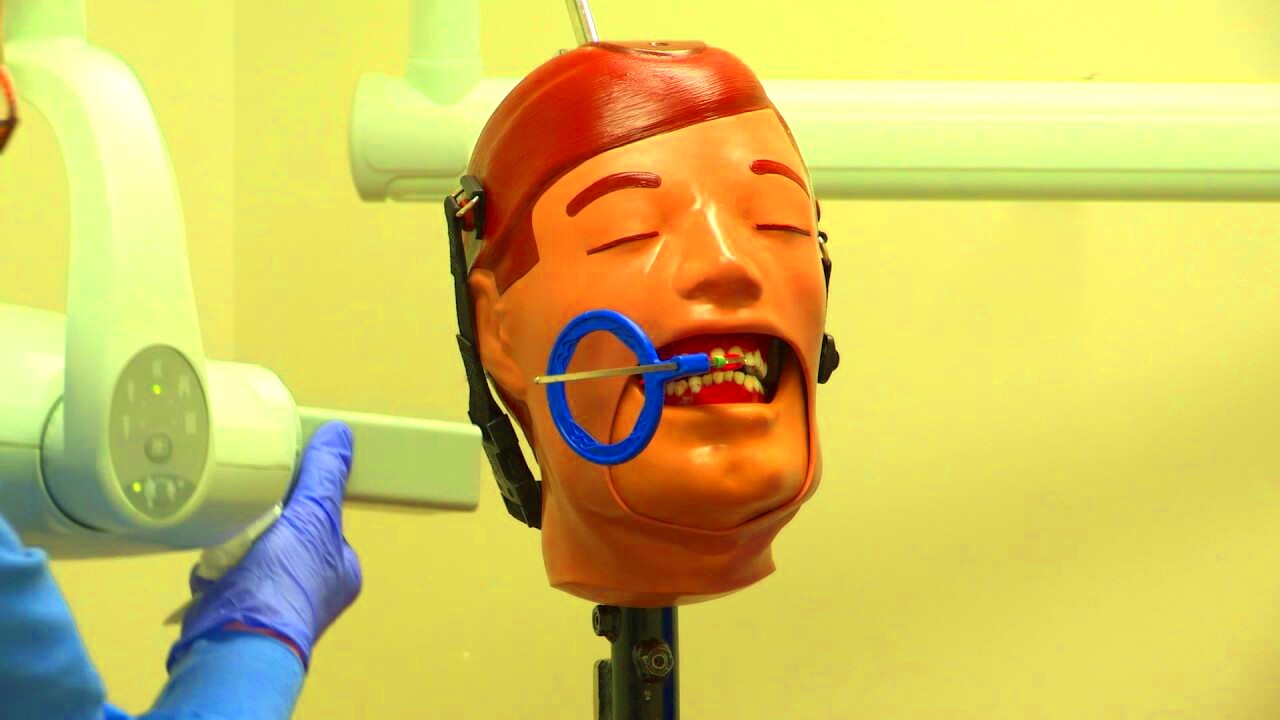
- Proper positioning of the patient: Ensuring that the patient is correctly positioned is vital for getting clear and accurate images. The dentist or dental hygienist will guide the patient to ensure they are in the right position.
- Proper technique and equipment: Dentists and dental staff should use high-quality equipment and proper imaging techniques to get the best results. This includes using the right exposure settings to reduce the risk of overexposure.
- Minimize radiation exposure: Dental professionals must adhere to the ALARA principle (As Low As Reasonably Achievable), which focuses on minimizing radiation exposure to both the patient and dental staff.
- Regular equipment calibration: The equipment used for intraoral imaging should be regularly checked and calibrated to ensure it is functioning correctly and producing accurate images.
By following these guidelines, both the dentist and patient can be assured that the intraoral images are accurate, safe, and used only when necessary to monitor oral health.
Also Read This: Enhancing Your Marketing Campaigns with Photos from Imago Images
Benefits of Regular Intraoral Imaging
Regular intraoral imaging offers several benefits that play a vital role in maintaining long-term oral health. While these images are commonly associated with detecting cavities, their value extends beyond that to provide a comprehensive view of a patient's overall dental condition. By scheduling routine imaging, dentists can monitor changes in oral health, spot potential problems early, and prevent more serious issues down the line.
Here are the key benefits of regular intraoral imaging:
- Early detection of issues: Regular intraoral imaging helps catch problems early, from cavities and gum disease to more complex issues like tooth infections or bone loss. Early detection often leads to simpler and more cost-effective treatments.
- Comprehensive monitoring: These images allow the dentist to track changes in the teeth, gums, and jaw structure over time. This is especially helpful in managing conditions like periodontal disease, where regular monitoring can guide treatment adjustments.
- Improved treatment planning: With detailed images, dentists can create more precise and personalized treatment plans. Whether it's a filling, root canal, or orthodontic work, accurate imaging helps ensure the right approach for each patient.
- Preventive care: Regular imaging is an excellent tool for preventive care. By identifying problems before they become severe, patients can avoid painful procedures and reduce the risk of costly dental treatments.
- Peace of mind: Knowing that a dentist is regularly checking for hidden issues can offer patients peace of mind, ensuring they are taking proactive steps to preserve their oral health.
In short, regular intraoral imaging helps keep your dental health on track, preventing problems from escalating and providing better overall care.
Also Read This: How Imago Images Keeps Up with Current Design and Visual Trends
Common Misconceptions About Intraoral Image Frequency
Despite the many benefits of intraoral imaging, there are several misconceptions that can lead to confusion regarding how often these images should be taken. Many people are uncertain about the need for regular imaging or may be concerned about the safety of radiation exposure. Let’s clear up some of the most common misunderstandings about intraoral image frequency.
Here are a few myths and facts about intraoral imaging:
- Myth 1: "Intraoral images are only needed when there’s a problem."
Fact: Intraoral images are essential even when there are no symptoms. They allow dentists to detect issues that may not be visible during a regular check-up, such as cavities between teeth or bone loss, preventing them from worsening.
- Myth 2: "Intraoral imaging exposes you to high levels of radiation."
Fact: Dental X-rays use very low levels of radiation. With modern technology and safety protocols like lead aprons, the amount of radiation a patient is exposed to is minimal and safe.
- Myth 3: "A complete series of intraoral images is needed every time you visit the dentist."
Fact: A full series of intraoral images is not needed at every visit. The frequency is determined based on your dental history and current oral health. Most adults with healthy teeth might only need a complete series every 3 to 5 years.
- Myth 4: "You should only have imaging done when you experience pain."
Fact: Dental issues often develop without symptoms, so regular imaging is key to catching problems early. By the time pain occurs, the issue may be more severe and require more invasive treatment.
Understanding the truth behind these myths can help patients feel more confident and informed about the role of intraoral imaging in their dental care routine.
Also Read This: In-Depth Reviews and Insights from Long-Time Imago Images Users
FAQ: Intraoral Images Frequency and Best Practices
When it comes to intraoral imaging, many patients have questions about how often these images should be taken, what the best practices are, and how the process works. To help clarify, we've compiled some of the most frequently asked questions about intraoral image frequency and best practices.
How often should I get intraoral images?
The frequency of intraoral imaging depends on several factors, including your age, oral health, and risk factors. Generally, adults with healthy teeth may need a complete series every 3 to 5 years. However, individuals with a history of dental issues or those at higher risk for oral health problems may require imaging more frequently, such as once every 1 to 2 years.
Are intraoral images safe?
Yes, intraoral imaging is safe. The amount of radiation used is very low, and modern equipment ensures that the exposure is minimal. Additionally, patients are protected with lead aprons and thyroid collars to further minimize radiation exposure.
Why do I need intraoral images if I’m not experiencing pain?
Intraoral images help detect dental problems before they cause pain or become noticeable. Conditions like cavities, gum disease, and bone loss often develop without symptoms. Regular imaging allows for early intervention, preventing these issues from becoming more serious and costly to treat.
Can intraoral images help with orthodontic treatment?
Yes, intraoral images are important in orthodontic treatment. They help dentists monitor the position of teeth, evaluate the bone structure, and check for any potential complications. By having detailed images, the dentist can plan more precise and effective treatment.
Are there any side effects from taking intraoral images?
Intraoral imaging has no side effects when used properly. The radiation exposure is minimal and regulated to ensure safety. If you are pregnant or have concerns about radiation, make sure to inform your dentist, who can take additional precautions or delay imaging if necessary.
By understanding these frequently asked questions, patients can feel more comfortable and informed about their dental care and the role that intraoral imaging plays in it.
Conclusion: Maintaining Oral Health with Intraoral Imaging
Intraoral imaging is a powerful tool that plays a crucial role in maintaining oral health. Regular imaging provides detailed insights into the health of your teeth, gums, and surrounding structures, helping dentists detect potential issues early. Whether it’s identifying cavities, monitoring gum health, or spotting hidden problems that aren’t visible to the naked eye, intraoral images ensure that problems are caught before they worsen.
By incorporating regular intraoral imaging into your dental care routine, you can proactively manage your oral health and prevent serious conditions down the road. The early detection of issues often leads to simpler, more affordable treatments, while also ensuring that any concerns are addressed promptly. In addition, the advancements in imaging technology have made the process quicker, safer, and more comfortable than ever before.
Ultimately, the goal is to maintain a healthy mouth for a lifetime. Intraoral imaging is a key part of that effort, enabling you and your dentist to stay on top of your oral health and keep your smile bright and healthy for years to come.

 admin
admin


