Ultrasound pictures are very important in medicine because they allow professionals to see what is inside a patient’s body. Sound waves are employed in these procedures so that organs, tissues and even blood movement can be observed as they happen. The ability to interpret these images can help diagnose different diseases including pregnancy management and heart conditions monitoring.In medicine, ultrasonic images make a huge difference as they enable doctors to see every single component within a human body. Sound waves work by producing live pictures of organs such as kidneys or lungs along with tissue strands or arteries supplying nutrients to different parts of your body. Analyzing these pictures could lead towards establishing several things like food management during pregnancy or looking at one’s own heart state.
The shades of gray you see represent different types of tissues. For instance: this is usually found throughout the body, especially in regions that support life. It can be noticed very easily on pregnant women’s babies in their wombs because these areas are called as placentas.In a way, ultrasound images can be considered as a new kind of photographs since they do not show the light like in pictures but sound waves are used to create such representations of internal organs. While looking at an ultrasound image you may notice that it looks somewhat different from a usual photograph; instead of capturing light ultrasonic sounds allow visualizing human body’s depths in quite an unusual manner. On such pictures various shades of gray represent different kinds of tissues. For instance:
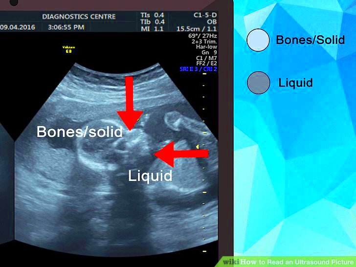
- White areas indicate denser tissues, like bone.
- Dark areas represent fluid-filled spaces, such as cysts.
- Gray areas are typically soft tissues.
It could be quite useful for one to grasp these fundamentals in order to understand ultrasound images better.
Types of Ultrasound Images
Ultrasound imaging comes in various forms that cater for different purposes. Below are some of the commonly known kinds:
- 2D Ultrasound: The standard type, producing flat images that show structures in a two-dimensional view.
- 3D Ultrasound: This provides a three-dimensional view of structures, often used in obstetrics to visualize the fetus.
- 4D Ultrasound: Similar to 3D but adds motion, allowing for real-time visualization, particularly useful in monitoring fetal development.
- Doppler Ultrasound: This type measures blood flow through vessels, helping diagnose issues like clots or arterial blockages.
For different kinds of ailments, every one of these variations has its own advantages and therefore it is very important to select the appropriate one.
Also Read This: Pixelating Images for Privacy and Style
How Ultrasound Works
Disentangling the mechanics of ultrasound can simplify matters. This is a concise description of the process:
- Sound Waves: Ultrasound uses high-frequency sound waves, which are above the range of human hearing.
- Transducer: A device called a transducer sends these sound waves into the body and picks up the echoes that bounce back.
- Echoes: The echoes return to the transducer, which then converts them into electrical signals.
- Image Creation: A computer processes these signals to create images displayed on a monitor.
The fact that ultrasound doesn’t involve any invasion of the body and radiations that are harmful can be considered one of its foremost benefits.
Also Read This: How to Create an Event on LinkedIn Organizing and Promoting Events through LinkedIn
Reading the Basics of Ultrasound Images
At first glance, reading ultrasound images might appear overwhelming, however, it is something anyone can learn to interpret much easier through practice. The initial move should be making yourself familiar with their general looks. Typically they have various tones of grey indicating diverse body tissues. Hence, it is important to comprehend what those shades stand for.
Here are a few things you should bear in mind:
- Hyperechoic: Areas that appear bright white; often indicate dense structures like bones or calcifications.
- Hypoechoic: Darker areas that suggest fluid-filled spaces or soft tissues.
- Anechoic: Completely black areas; typically indicate fluid without any internal echoes, like cysts.
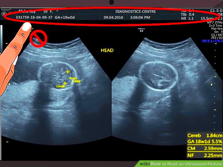
Understanding how an image is oriented is also essential. Most of the time, the patient's right is represented on the left side of the image. This is particularly important when you have to talk about your findings with a doctor. However, be patient with yourself; it takes time to learn these images and don’t be afraid to raise any concerns or ask questions if you do not understand something.
Also Read This: Exploring Revenue Opportunities: Making Money from 123RF Images
Identifying Key Features in Ultrasound Images
Once you have gained some skills in handling the fundamentals, you can start identifying main components inside ultrasound pictures. This will allow you to have a better understanding of what you are investigating. Important characteristics include the following:
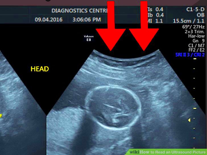
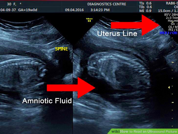
- Gestational Sac: In pregnancy ultrasounds, look for the gestational sac, which appears as a dark area indicating the developing fetus.
- Fetal Heartbeat: A flickering spot within the gestational sac, often identifiable around 6-7 weeks of pregnancy.
- Organs: Familiarize yourself with the shapes of organs. For example, the liver appears as a bright structure in the upper right abdomen.
Utilizing a diagram with labels may are also advantageous. With the aid of an organ chart for instance showing how those organs will appear in a sonar (ultrasound) scan you’ll be able to learn effectively. Below is a brief comparison:
| Organ | Appearance |
|---|---|
| Liver | Bright, slightly textured surface |
| Kidneys | Bean-shaped with a distinct cortex |
| Gallbladder | Dark, oval shape |
Once you habituate yourself to these features, your understanding will deepen and interpretation of ultrasound images will become more effortless for you.
Also Read This: Elevate Your Presentation Designs with 123RF
Common Misinterpretations of Ultrasound Images
When it comes to interpreting images from ultrasound scans, exceptions should be avoided because they might generate false impressions. Here are some common misconstructions and tips on how you can prevent them:
- Confusing Shadows: Dark shadows may be mistaken for abnormalities. However, they can also result from normal anatomy.
- Size Miscalculations: The perceived size of structures can be misleading due to the angle of the ultrasound beam.
- Fetal Position: The position of the fetus can affect the visibility of certain features, leading to assumptions about growth or health.
To minimize these misunderstandings, it is important to trust the qualified healthcare professional for interpreting. They have the education and experience to distinguish between what is normal and what is not. If ever in doubt about an image, do not hesitate to seek clarification. Key to our understanding of what we see but professional input is important for right assessments.
Also Read This: How to Create a Slide Post on LinkedIn with Simple Steps
Practical Tips for Viewing Ultrasound Images
Seeing ultrasound images might seem like a daunting task initially. However, by putting into practice some of the suggestions given below, the whole experience would become easy. These recommendations will also assist patients and curious persons on how to better understand them.
Following are some useful tips:
- Ask for Explanations: Don’t hesitate to ask your healthcare provider to explain what you are looking at. They can help you understand the images and what to look for.
- Use a Reference: Having a reference guide or a labeled diagram can make it easier to identify various organs and structures on the ultrasound image.
- Stay Calm: If you’re the patient, try to relax during the ultrasound. Tension can affect how well the images turn out.
- Consider the Context: Remember that the ultrasound is just one part of the diagnostic process. Context from your medical history and symptoms is essential.
In addition, it is beneficial to analyze the pictures together at home with an informed individual. Having another set of eyes may provide a new viewpoint and extra understanding creating a more joint endeavor.
Also Read This: DIY Nail Art Techniques at Home
Frequently Asked Questions
Ultrasound images prompt numerous inquiries which include but they are not limited to. Below are available answers on some frequently asked questions:
- What does a black area mean? A black area on an ultrasound image typically indicates a fluid-filled space, such as a cyst.
- Can ultrasound images show everything? While ultrasound is a valuable tool, it cannot visualize all conditions. Additional imaging methods may be necessary.
- How long does it take to get results? Most ultrasound results are available quickly, often within a day or two. However, your doctor will discuss the findings during your follow-up appointment.
In the light of these FAQs, it’s evident that patient- physician communication is vital. However, if you have any other questions, feel free to ask your doctor or technician for more information.
Conclusion
Despite the fact that you may encounter a lot of people who are not so much into medical imaging, the more you take time to learn about reading ultrasound images, the more benefit you will get from it. Understanding ultrasound images is important in letting one understand about his/her health status and gaining confidence in that regard. Also keep in mind, if you have questions, it is okay because this always gives room for further discussion with qualified persons as they can answer better than us.
For now, it looks that way. Although you may be undertaking in anticipation of a child or keeping track of an illness; ultrasound offers an unparalleled perspective on the interior of our bodies. Welcome this process as a chance to become increasingly knowledgeable about it and don’t be afraid to ask for assistance if necessary. There’s no reason why these practical tips and pieces of advice won’t help you to decode the ultrasound pictures!
 admin
admin