Heat maps are powerful visual tools used to represent data density over a small two-dimensional region. In terms of CT imaging, heat maps help radiologists and healthcare professionals identify patterns and anomalies inside the images. They transform complex data into an easy-to-understand form showing regions of interest visualized by means of the intensity of signals. This may be essential for diagnosing conditions, monitoring how effective treatments were as well as when planning surgical interventions.
In this manner, healthcare workers can interactively assimilate vital information and take improved steps in decision-making. One example of such implementation is the use of heat maps that would indicate which areas of a tissue are most affected by a given disease and thus allow the doctors to concentrate their analysis on acute areas.
Gathering the Required Tools for Drawing Heat Maps
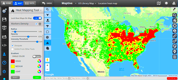
Thus, you must collect the appropriate instruments before beginning the process of developing temperature maps. A simple inventory of necessary things includes:
- Software: Programs like MATLAB, Python (with libraries such as Matplotlib), or dedicated imaging software.
- CT Image Files: Ensure you have access to high-quality CT images in a compatible format, such as DICOM.
- Data Analysis Tools: Familiarity with data analysis techniques will help you manipulate and visualize the data effectively.
- Color Palette Selection: Choose a color scheme that effectively represents the data's intensity and is easy to interpret.
Remember these tools can be used to make the process easier and simpler.
Step-by-Step Guide to Preparing CT Images
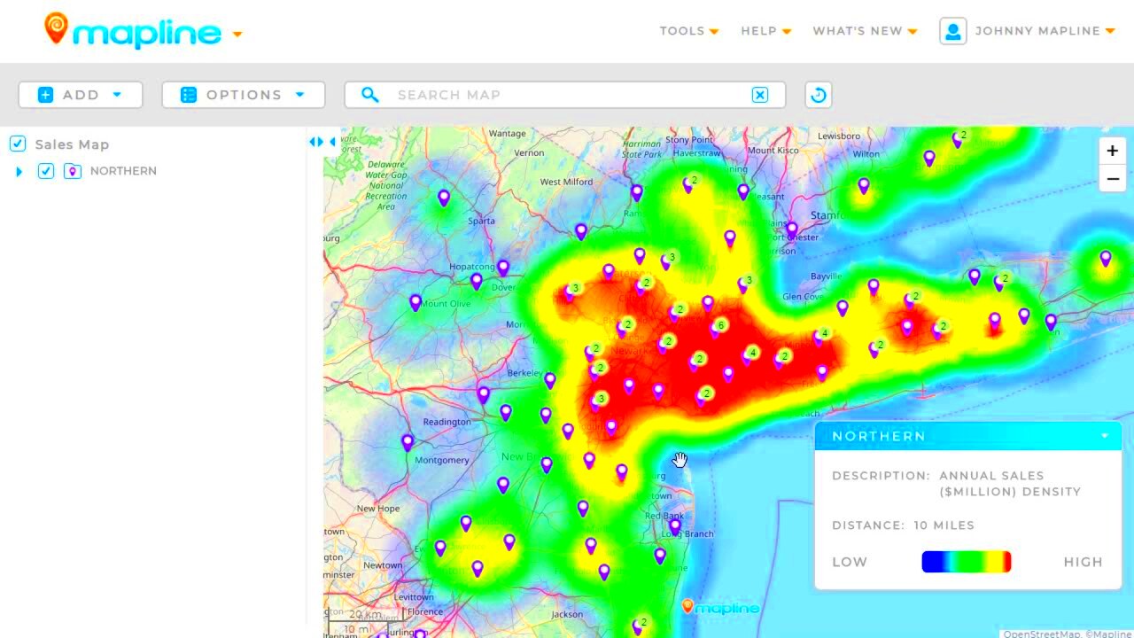
Your CT images should be prepared carefully for the heat map creation as this might lead to significant changes in the outcomes. Make sure that you consider the following tips if you want to make sure that your images are appropriate:
- Import the CT Images: Load the DICOM files into your chosen software.
- Preprocess the Images: Clean the images by removing noise or artifacts that could affect the analysis.
- Standardize Image Sizes: Ensure all images are of the same resolution and dimensions for accurate comparison.
- Apply Necessary Filters: Depending on your analysis needs, apply filters to enhance specific features within the images.
- Segment the Images: Identify and isolate areas of interest. This might involve using software tools to outline tumors or other relevant structures.
- Export the Processed Images: Save your images in a suitable format for heat map generation.
So once you have doneyour CTpictrue accordingthe way it was said, you’ll be able produce productive thermographs which will bring forth the significance ofthe data.
Creating Heat Maps Using Software Tools
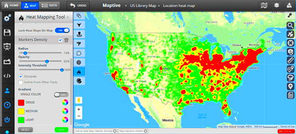
Once you have patched up your CT pictures, we are now in a position to make heat maps. Different software tools can help you achieve this, each with its unique features and advantages. Here’s how to create heat maps using some common tools:
- Using Python: If you're comfortable with coding, Python offers libraries like Matplotlib and Seaborn that are perfect for generating heat maps. You can load your data, customize the color schemes, and visualize the heat map with just a few lines of code.
- Using MATLAB: MATLAB has built-in functions to create heat maps. Simply input your data, choose your color map, and generate the visual. It's especially useful for matrix data.
- Dedicated Imaging Software: Tools like OsiriX or ImageJ are designed specifically for medical imaging. They often have user-friendly interfaces where you can import your CT images, apply color maps, and instantly visualize the heat map.
Nevertheless, typically the fundamental phases involve using any instrument of your preference:
- Loading the processed CT images.
- Selecting the area of interest for heat map generation.
- Adjusting color scales to represent data intensity accurately.
- Saving or exporting the heat map for further analysis.
Generating heat maps can be an easy task when using the appropriate software therefore you must take your time to examine what the tool of your choice has to offer.
Interpreting the Results of Your Heat Map
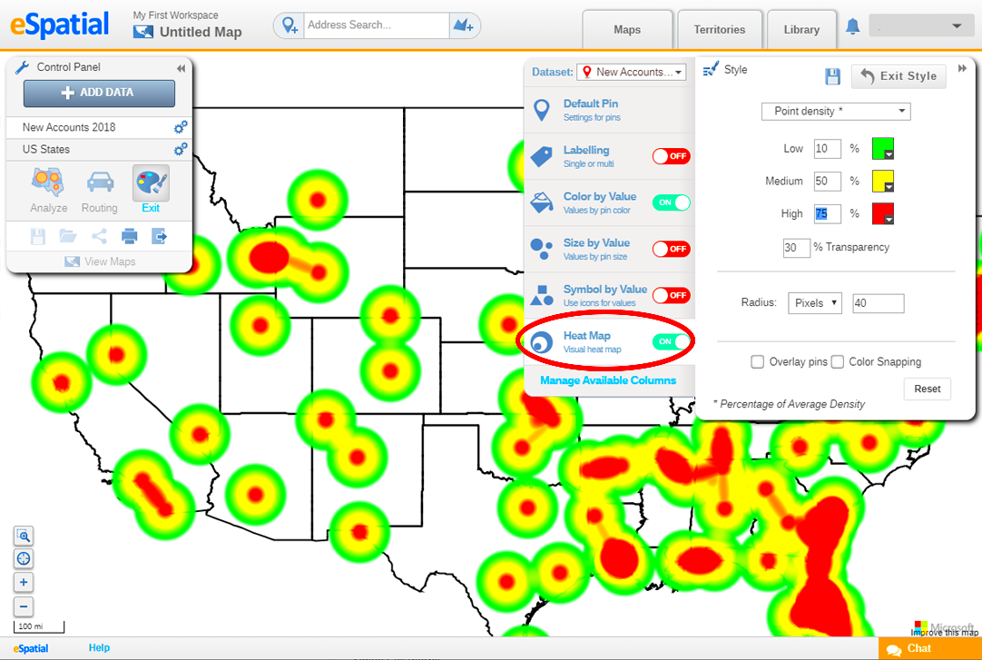
Once your heat map is created, it becomes critical to interpret results properly. Usually interpretation mainly depend on the color scheme where by warm colors symbolize higher infrequency while cool colors indicate lesser frequency. Here’s how to make sense of what you see:
- Identify Hotspots: Look for areas with intense colors; these represent regions with higher activity or concentration of signals. In medical imaging, these could indicate tumors or areas of inflammation.
- Analyze Patterns: Observe the overall patterns. Are there clusters of activity? This might suggest underlying conditions that need further investigation.
- Compare with Clinical Data: Always cross-reference heat map findings with clinical data or other imaging results. This will provide a more comprehensive view of the patient's condition.
Bear in mind that mixing technical capabilities and clinical information is necessary for interpreting heating diagrams. Moreover, it is prudent to share your observations with a radiologist or any medical personnel in order to reach precise decisions.
Common Mistakes to Avoid When Drawing Heat Maps
It can be an easy thing to create heat maps, however there are standard mistakes that make them inaccurate or misrepresentative. These are common things to keep in mind:
- Ignoring Image Quality: Low-quality images can lead to poor heat map outputs. Always start with high-resolution CT images to ensure accuracy.
- Choosing Inappropriate Color Scales: Using colors that are too similar can obscure important data. Make sure your color palette effectively distinguishes between varying intensities.
- Overcomplicating the Data: Adding too many variables can make the heat map cluttered and confusing. Focus on key areas of interest for clarity.
- Neglecting Calibration: Failing to calibrate the software correctly can lead to skewed results. Ensure all settings are accurate before generating heat maps.
- Forgetting to Validate Findings: Always cross-check heat map results with other data sources or imaging techniques. Validation is crucial for drawing reliable conclusions.
These common mistakes should be avoided in order to produce better and more meaningful heat maps which will eventually strengthen the analytical and decision making process.
Practical Applications of Heat Maps in CT Imaging
Топлото дакота не се само визуелизација. Веројатно во различни медицински примени многу помагаат, особено во снимањето на CT. Со претворање на сложените податоци во визуелни формати, топли мапи помагаат на медицинските лица да донесуваат намерни одлуки. Такви се некои практични апликации:
- Identifying Tumors: Heat maps can highlight areas of abnormal tissue density, aiding in the early detection and diagnosis of tumors. This can be particularly beneficial in oncology.
- Monitoring Treatment Progress: After treatment, heat maps can visually represent changes in tissue density, helping doctors assess the effectiveness of therapies over time.
- Guiding Surgical Planning: Surgeons can use heat maps to identify critical areas to target during procedures, improving precision and outcomes.
- Studying Organ Function: Heat maps can illustrate the functionality of different organs by showing how they absorb contrast agents during imaging, aiding in diagnosing conditions like ischemia or infarction.
In summary, heat map applications in CT imaging go way beyond simple visualization; they give meaningful information that can help in improving the quality of patient care and results from treatment.
Frequently Asked Questions about Heat Maps for CT Images
Diverse individuals pose inquiries regarding heat maps, particularly in relation to CT imaging. Below are some often raised queries that can assist in elucidating usual concerns:
| Question | Answer |
|---|---|
| What is a heat map? | A heat map is a data visualization technique that uses colors to represent the intensity of data in a specific area. |
| How are heat maps created from CT images? | Heat maps are generated by processing CT images through software that analyzes data intensity and visualizes it using color gradients. |
| Are heat maps reliable for diagnosis? | When interpreted correctly and used alongside other clinical data, heat maps can be reliable tools for diagnosis. |
| Can I create heat maps without programming skills? | Yes! Many imaging software options provide user-friendly interfaces that allow you to create heat maps without programming. |
If you have any more inquiries, please feel free to contact an expert or conduct additional research in order to obtain correct information.
Conclusion on the Significance of Heat Maps in Medical Imaging
CT scans often rely on heat maps that help in providing an effective way of mapping complex data so that they can be used by those dealing with patients to understand what to do or what has been done. They pinpoint problematic spots, follow the progress of treatment procedures and support surgical planning leading to increased outcomes.
Not only do the doctors get help in diagnosing, raw data can be changed into meaningful diagrams. More efficient contact points between patients and medical professionals is what this promotes. The incorporation of heat maps in healthcare services will become even more common with advancements in technology; hence enabling improved results and better health care options.
Understanding and using heat maps can ultimately enable both healthcare practitioners and patients which in turn makes them significant instruments in present day medical imaging.
 admin
admin


